Core facilities Uni Heidelberg EMCF
The Electron Microscopy Core Facility (EMCF) provides expertise in electron microscopy imaging, for everyone who wants to examine their specimen on a sub-cellular level. We work together with research groups from the whole Heidelberg campus to help find the most suitable sample preparation and imaging technique to answer various biological questions related to ultra structure.
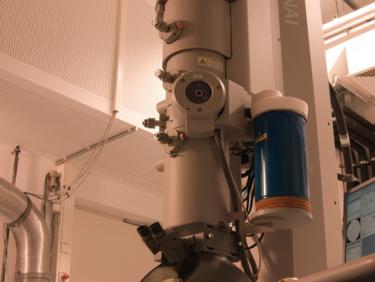
To be able to image samples in the electron microscope the specimen has to be prepared to withstand vacuum conditions and to give contrast in an electron beam. The necessary preparations can be done by EMCF personnel, but it is also possible for you to learn the methods and work independently using our equipment. For imaging a couple of different electron microscopes are available at the facility, and they are used for different purposes. With the scanning electron microscope, we can image surface structures of cells and tissues, which is useful both for fine structures on single cells as well as for looking at details from the surface of tissues or whole (mm size) organisms. To investigate intracellular structures, the samples have to be resin embedded and sectioned in ultrathin slices with a thickness of about 70 to 200nm. In the transmission electron microscope, the structures within the sections can be examined and all kinds of intracellular structures from large vacuoles to small ribosomes can be visualized. In addition, we also have a microscope with a higher accelerating voltage (200kV), which enables taking images of the section at different tilt angles and with a tilt series a 3D reconstruction of the fine structures within the section can be obtained. This makes it possible to look at how small structures are spatially organised in the sectioned slice of the specimen. When a 3D organisation of a larger volume is of interest using serial sections of a sample is an option. This can be done both with transmission electron microscopy or with scanning electron microscopy - array tomography. For extracted protein complexes or extracted cellular components we use negative stain and transmission electron microscopy to gain insights in their structure. In summary, we offer EM techniques covering from surfaces of tissues to fine structure of protein complexes.

To find out more about the EMCF please visit our home page.
To start an EM project contact us via email: emcf@uni-heidelberg.de


